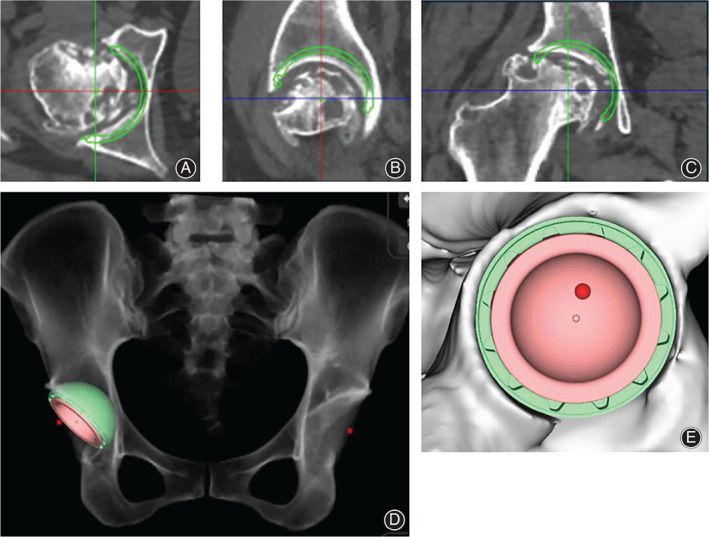
Fig. 1.

CT‐based planning for acetabular component. (A) Simulation of acetabular component in axial view; (B) Simulation of acetabular component in sagittal view; (C) Simulation of acetabular component in coronal view; (D) Simulation of postoperative X‐ray; (E) Simulation of peripheral cup coverage. The green circle indicates where the stem is implanted in simulation. The red dot indicates the position of original hip center. In this case, the cup was planned at 20° anteversion, 45° inclination, and the cup coverage was 92%.
