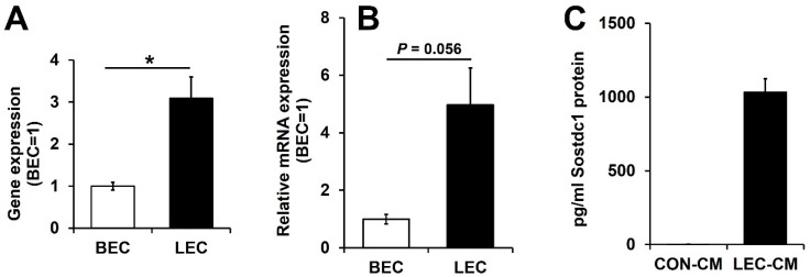
Figure 1.
Sostdc1 is highly expressed in lymphatic endothelial cells compared with blood vascular endothelial cells. (A,B) The expression of Sostdc1 in matched pairs of human LECs and BECs was determined by microarray (A) and by qRT-PCR (B). (C) Sostdc1 protein levels were measured by ELISA in conditioned media of control (CON-CM) and conditioned media of LECs (LEC-CM). Results are expressed as mean ± standard error of the mean (SEM). Data were analyzed using a two-tailed unpaired t-test. * p < 0.05 compared to the control group.

