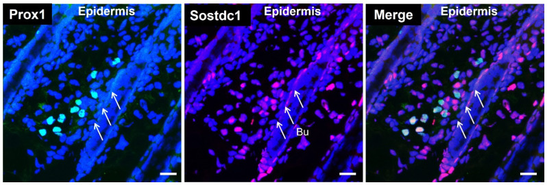
Figure 2.
Lymphatic vessels express Sostdc1. Immunofluorescent staining of 10-µm frozen sections of back skin (anagen phase, postnatal day 8) for Prox1 (lymphatic-specific transcription factor, green) and Sostdc1 (red). Nuclear staining with Hoechst 33342 (blue). White arrows indicate the bulge area (Bu). Scale bars: 20 μm.

