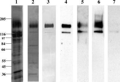
FIG. 1.
Analysis of pool I Cry1Ac and Cry1Aa toxin-binding proteins. One microgram of protein was loaded in each lane, and the proteins were separated on 8% polyacrylamide SDS-PAGE gels. For immunoblotting, toxin overlay assays, and lectin blotting, the proteins were transferred to an Immobilon-P membrane. Lane 1, silver-stained CHAPS-solubilized BBMV; lane 2, silver-stained pool I MonoQ fraction; lane 3, soybean agglutinin lectin blot detection of glycosylated proteins in pool I; lane 4, Cry1Aa toxin binding to pool I proteins; lane 5, Cry1Aa toxin binding to pool I proteins in the presence of 100 mM GalNAc; lane 6, Cry1Ac toxin binding to pool I proteins; lane 7, Cry1Ac toxin binding in the presence of 100 mM GalNAc. The positions of molecular mass markers (in kilodaltons) are indicated on the left.

