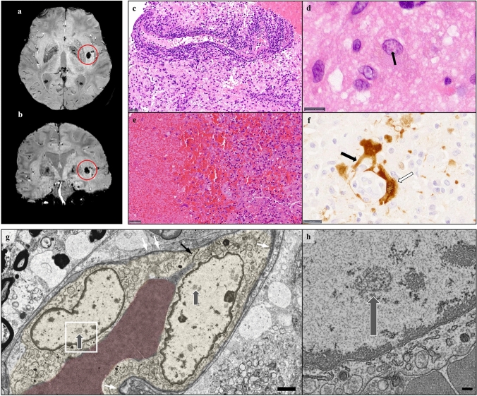
Fig. 1.
a, b MRI susceptibility weighted imaging (SWI) reveals a subinsular hemorrhage (red circles) 26 days after onset of symptoms. c, d Hematoxylin and eosin (HE) of a biopsy from the left caudate nucleus shows strong lymphocytic infiltrates (c, scale bar 20 μm) and astrocytes with eosinophilic intranuclear inclusion bodies (d, scale bar 10 μm). e, f In autoptic material, the hemorrhage with surrounding macrophages was seen (e, HE, scale bar 50 µm). Strikingly, a positive immunoreaction for BoDV-1 was detected in components of vessels (white arrow) and endfeet of astrocytes (black arrow) (f, scale bar 20 µm). g Electron microscopy shows two infected endothelial cells (yellow) with replication centers (grey arrows). For orientation, tight junctions (black arrow) and basal membranes (white arrows) are marked (scale bar 2 µm). h Higher magnification view of g (area within rectangle) displays detailed architecture of a viral replication center (arrow; scale bar 200 nm)

