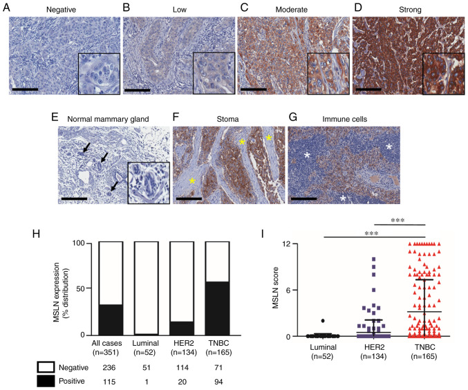
Figure 1.
Expression of MSLN in human breast cancer tissue samples. Representative images of MSLN expression levels in breast cancer tissues ranging from (A) negative, (B) low, (C) moderate and (D) strong staining. (E) Normal lobules (black arrow) showed no MSLN expression. Negative staining of MSLN in (F) stromal cells (yellow asterisk) and (G) immune cells (white asterisk). (H) Proportion of MSLN expressing samples in full cohort and stratified subtypes. (I) Mean MSLN score stratified by subtypes of breast cancer. Original magnification, 200×; scale bars=200 µm. ***P<0.001. MSLN, mesothelin.

