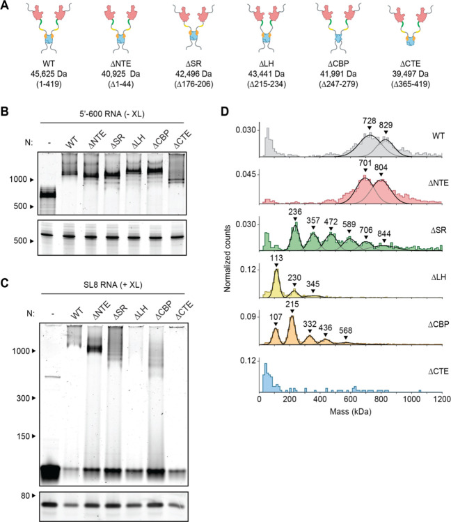
Figure 3. Disordered regions contribute to vRNP formation.
(A) Schematic of wild-type (WT) N protein and deletion mutants, as described in the text. Mass is that of monomeric N protein. (B) 15 μM N protein mutants were mixed with 256 ng/μl 5’−600 RNA and analyzed by native (top) and denaturing (bottom) gel electrophoresis. (C) 20 μM N protein mutants were mixed with 256 ng/μl SL8 RNA and analyzed by native (top) and denaturing (bottom) gel electrophoresis. SL8 ribonucleoprotein complexes were crosslinked prior to analysis. (D) Mass photometry analysis of crosslinked N protein mutants (20 μM) bound to SL8 RNA (256 ng/μl). Representative of at least two independent experiments (table S1).

