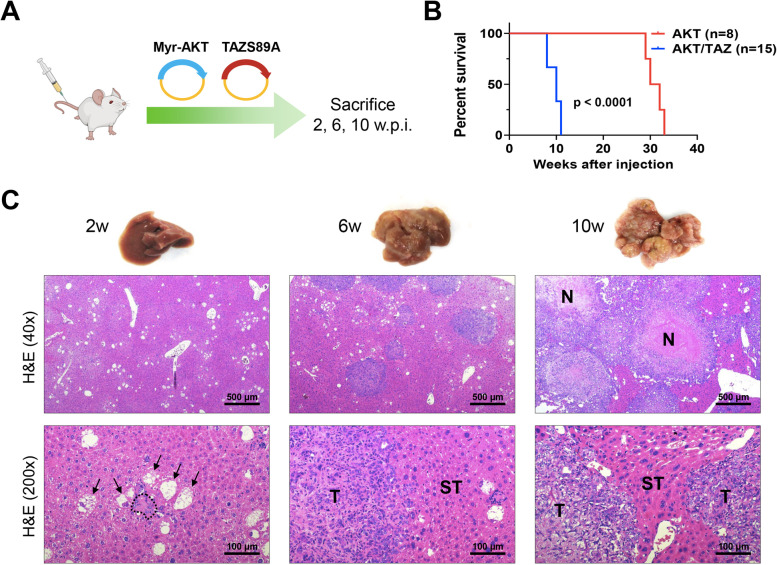
Fig. 2.
Overexpression of constitutively activated TAZ (TAZS89A) synergizes with AKT to induce intrahepatic cholangiocarcinoma development in mice. (A) Scheme of the experiments conducted. FVB/N mice were hydrodynamically injected with a myristoylated/activated form of AKT (Myr-AKT) and an activated form of TAZ (TAZS89A; n=25; these mice are referred to as AKT/TAZ mice). Eight mice were injected only with Myr-AKT and are referred to as AKT mice empty vector-injected mice. Five mice were sacrificed at 2 weeks post-injection and additional five mice at 6 weeks post-injection for the assessment of early lesions. Fifteen AKT/TAZ-injected mice were monitored and sacrificed 10 weeks post-injection when a high tumor burden occurred. (B) Survival curves of AKT and AKT/TAZ mice. (C) Macroscopic and microscopic images of AKT/TAZ mouse livers. Two weeks post-injections, single cells and clusters of a few cells are appreciable in the AKT/TAZ livers. Besides normal hepatocytes, large cells owing to lipid accumulation (indicated by arrows) and small cells with scarce cytoplasm (surrounded by dots) can be easily detected. By six weeks post-injection, most giant cells had disappeared, and tumors of cholangiocellular features developed the liver parenchyma. By 10 weeks post-injection, tumors had progressed, occupying most of the liver surface. Signs of biological aggressiveness, such as large areas of necrosis (N), are visible. Original magnification: 40x and 200x; scale bar: 500 μm in 40x, 100 μm in 200x. Abbreviations: H&E, hematoxylin and eosin staining; w.p.i, weeks post-injection

