Fig. 4.
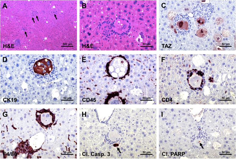
Early lesions developed in AKT/TAZ mice display an inflammatory infiltrate. (A) Two weeks after hydrodynamic injection of AKT and TAZ, numerous transfected cells (indicated by black arrows) were encircled by an inflammatory infiltrate. The transfected cells and the inflammatory cells could be better appreciated at higher magnification (B), with the inflammatory cells characterized by small spherical nuclei and forming a fence around the transfected cells. The transfected cells displayed elevated TAZ (C) and CK19 (D) immunoreactivity. The inflammatory cells consisted of lymphocytes, characterized by immunoreactivity for CD4 and CD45 markers (E, F), and macrophages (immunoreactivity for the F4/80 marker) (G). Some small cells encircled by the inflammatory reaction showed signs of apoptosis, as revealed by positive immunoreactivity of apoptotic bodies (indicated by black arrows) for cleaved Caspase 3 (H) and cleaved PARP (I), implying their destruction by the inflammatory response. Original magnification: 100x in (A), 400x in (B-I); scale bar: 200 μm in (A), 50 μm in (B-I).

