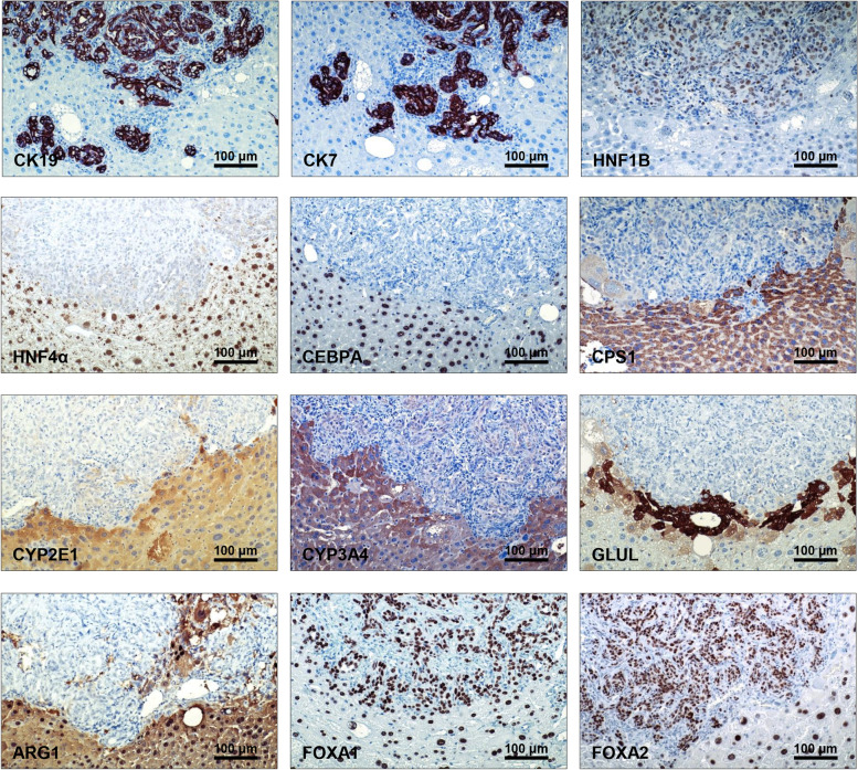
Fig. 6.
AKT/TAZ liver lesions exhibit molecular features of cholangiocarcinoma. Representative immunohistochemical patterns of an AKT/TAZ tumor showing positive immunoreactivity for cholangiocellular markers such as cytokeratin (CK) 7, CK19, and HNF1B, and weak/absent immunolabeling for hepatocellular markers, including hepatocyte nuclear factor (HNF)-4α, CCAAT/enhancer-binding protein (CEBP)-A, Carbamoylphosphat-Synthetase I (CPS1), Cytochrome P450 Family 2 Subfamily E Member 1 (CYP2E1), Cytochrome P450 3A4 (CYP3A4), glutamine synthetase (GLUL), and liver arginase (ARG1). The staining pictures are sections from the same tumor depicted in Fig. 4. Original magnification: 200x; scale bar: 100 μm

