Figure 7.
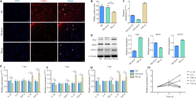
The effect of different expression levels of miR-181b on cerebral cell apoptosis and neuroinflammation in injured area of TBI mice.
(A) TUNEL staining showing the levels of cerebral cell apoptosis (arrows) in the three groups. There were fewer apoptotic cells (TUNEL-positive cells) in the TBI-up group than in the other two groups. Scale bars: 0.2 mm. (B) Quantification of TUNEL-positive cells. (C) Expression level of miR-181b 7 days after the transfection with lentivirus. (D) Protein expression of microglia markers, Arg1 and iNOS, as well as the transcriptional regulator, STAT3. (E) mRNA expression of inflammatory factors on days 1, 3, and 7 post TBI (I–III). Dynamic changes in inflammatory factors in TBI-up group (IV). Data are expressed as mean ± SD (n = 5 per group). *P < 0.05 (one-way analysis of variance followed by the least significant difference test). Arg1: Arginase-1; DAPI: 4′,6-diamidino-2-phenylindole; Dpi: days post injury; IL: interleukin; iNOS: inducible nitric oxide synthase; STAT3: signal transducer and activator of transcription 3; TBI: traumatic brain injury; TGF-β: transforming growth factor-β; TNF-α: tumor necrosis factor-α.

