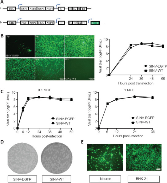
Figure 1.

The biological characteristics of the SINV-EGFP in vitro.
(A) Diagram of pSINV-EGFP (a) and pSINV-WT (b) genome structures. (B) The kinetics of virus production. Fluorescent images of BHK-21 cells after transfecting pSINV-EGFP and pSINV-WT at different time points. Fluorescence signals could be detected at 12 hpt and increased with time in the pSINV-EGFP group. No fluorescence was detected in the pSINV-WT group. Virus titers at different time points post-transfection were measured by plaque assay. (C) The single-step growth curves of both viruses. These viruses were collected and titered on BHK-21 cells at different time points post-infection. (D) Plaque sizes of both viruses. The sizes of the viruses were not significantly different. (E) Fluorescent images of cultured primary neurons and BHK-21 cells after infecting with SINV-EGFP. All cells were infected and expressed EGFP. C: Capsid; E3, E2, E1, 6K: structural protein; EGFP: enhanced green fluorescent protein; hpt: hours post-injection; MOI: multiplicity of infection; NSP1–4: nonstructural protein 1–4; UBC: ubiquitin C promoter.
