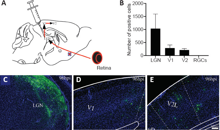
Figure 2.

Injection into the LGN of mice to characterize virus trans-synaptic properties.
(A) Diagram of circuits between retinal ganglion cells, LGN, V1 and V2. Data are expressed as mean ± SD. (B) Positive cells of different brain areas at 96 hours post-injection. (C–E) SINV-EGFP spread transsynaptically in the anterograde direction from injected sites to primary output V1 and secondary output V2 by 96 hpi. A large number of neurons were labeled in the LGN (C), and positive cells were also detected in V1 (D) and V2 (E). The experiments were repeated three times. EGFP: Enhanced green fluorescent protein; hpi: hours post-injection; LGN: lateral geniculate nucleus; RGCs: retinal ganglion cells; V1: visual cortex area 1; V2: visual cortex area 2.
