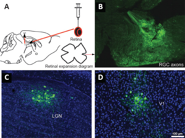
Figure 3.

Injection into the retina of mice to characterize the virus.
We injected 2 μL SINV-EGFP into the vitreous body of the eye of mice, and mice were sacrificed at 96 hpi. (A) Diagram of neural circuit between the RGCs of retina, the LGN and V1. (B) At 96 hpi of subretinal cells, we isolated and expanded retinal cells. RGC axons were labeled by SINV-EGFP. (C, D) Positive signals were detected in the LGN and V1, which indicates that the virus anterogradely spread in the neural circuit. The experiments were repeated three times. EGFP: Enhanced green fluorescent protein; hpi; hours post-injection; LGN: lateral geniculate nucleus; RGCs: retinal ganglion cells; V1: visual cortex area 1.
