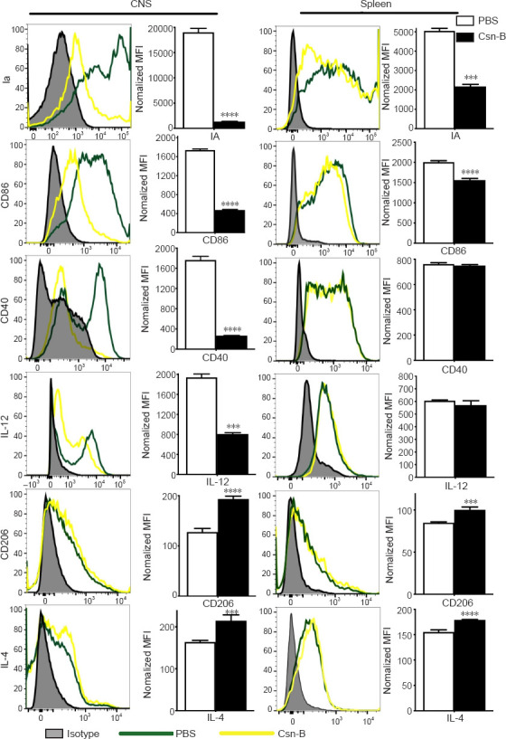
Figure 5.

Macrophages/microglia display an enhanced anti-inflammatory cell phenotype after cytosporone B (Csn-B) treatment.
After induction of experimental autoimmune encephalomyelitis, 10 mg/kg Csn-B or PBS was administered once per day from day 14 to 21 post-immunization. The mice were sacrificed on day 21. Infiltrating cells in the central nervous system (CNS) and splenocytes were isolated and stained with the pan-macrophage marker F4/80; M1 markers Ia, CD86, CD40, and IL-12; and the M2 markers CD206 and IL-4. The cells were analyzed using flow cytometry. Expression of these markers in the CNS and spleens was presented as the mean fluorescence intensity (MFI) (left), and the quantitative results are shown in the bar graphs (right). Data are expressed as means ± SEM. ***P < 0.005, ****P < 0.001 (Student’s t-test). Csn-B: Cytosporone B; IA: I region associated antigen; IL: interleukin; MOG35–55: myelin oligodendrocyte glycoprotein-35–55; PBS: phosphate buffer saline.
