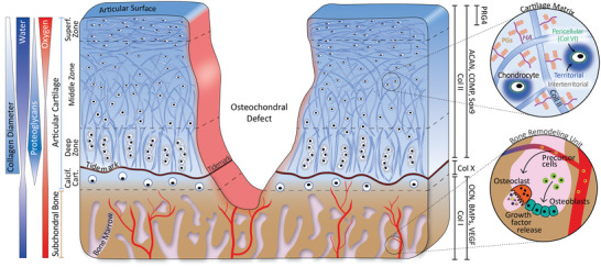
Figure 1.

Schematic cross‐sectional representation of the OC unit presenting an ICRS grade IV OC defect. The different regions (subchondral bone, calcified cartilage, deep zone, middle zone, and superficial zone) and gradients of the unit are presented on the left. The main markers of the different zones are presented on the right. These include collagen (Col) types II and X, aggrecan (ACAN), cartilage oligomeric matrix protein (COMP), Sox9 and proteoglycan 4 (PRG4) for articular cartilage; and Col I, osteocalcin (OCN), Bone Morphogenic Proteins (BMPs), and vascular endothelial growth factor (VEGF) for the subchondral bone. The two circles are enlarged views of the cartilaginous and bone tissues. In cartilage, chondrocytes are embedded in a matrix mainly composed of Col II, hyaluronic acid (HA), and proteoglycans (PGs). In the subchondral bone, osteoblasts and osteoclasts remodel the matrix.
