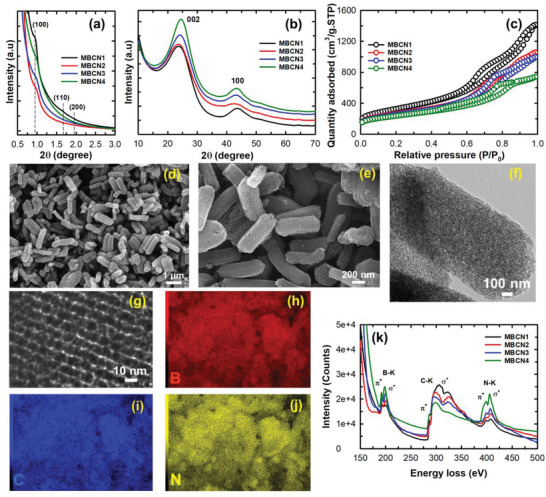
Figure 2.

a) Low‐angle and b) high‐angle powder X‐ray diffraction patterns. c) N2 adsorption–desorption isotherm. d,e) SEM images showing rod‐like morphology of MBCN1. f,g) HRTEM images showing an ordered porous structure in MBCN1 and the corresponding EDX mapping of h) boron, i) carbon, j) nitrogen of MBCN1 sample, and k) EELS spectra of MBCN samples.
