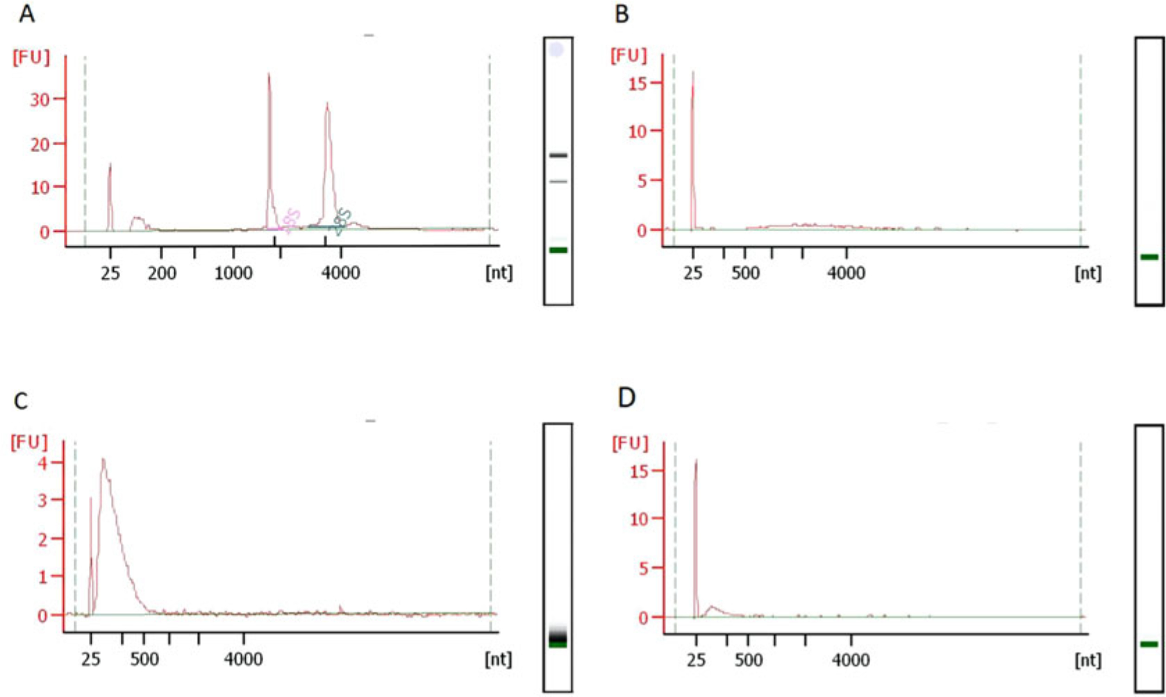
Fig. 3.

Typical RNA fragment size-distributions during RibOxi-seq. All QC in this figure was done using Bioanalyzer Nano 6000 chips. (a) Typical size distributions of total RNA extracted from HEK293T cells with RNA integrity number of 10. (b) After poly(A) enrichment twice, there should not be any major rRNA peaks left. (c) The RNA size distribution after benzonase fragmentation. (d) Size distribution after 3′-linker ligation. A size shift is not observable, most likely due to the fact that only a very small portion of the RNA was protected from oxidation by Nm and ligated with linkers
