
Fig. 4.
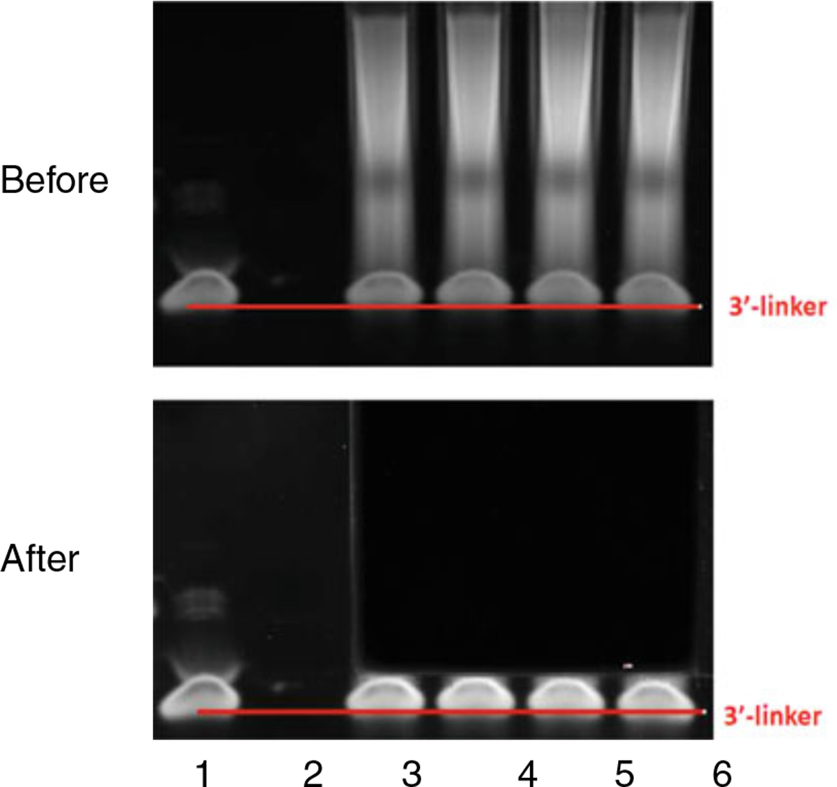
A typical PAGE gel pattern before and after cutting. Lane 1 shows RibOxi-seq 3′-linker. Lanes 3–6 contain reactions postligation. The goal is to recover RNAs and exclude free linkers. The top panel illustrates a typical electrophoresis. The bottom panel illustrates where we normally cut