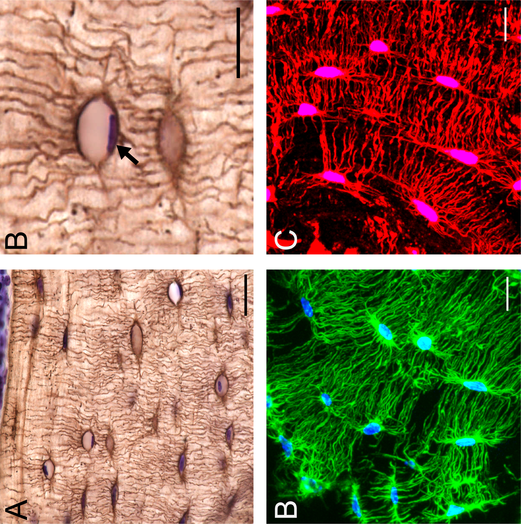
Figure 1:

Stains highlighting morphological changes in osteocytes associated with PLR. Silver nitrate stain on C57/BL6 mouse bone demonstrates the osteocyte cell network in the paraffin embedded cortical bone (A, scale bar = 20 μm) with cresyl violet counter stain for nuclei (purple). Enlarged region of section from A (B, scale bar = 10 μm) depicting an osteocyte containing a positively stained cresyl violet nuceli (black arrow). Fluorescent phalloidin (C, scale bar = 10 μm) staining of f-actin (green) and DAPI (blue), can be used to visualize the osteocyte cell network in 3D with frozen,OCT sectioning and confocal microscopy. Dil stain (red) (D, scale bar = 10 μm) intercalates into the hydrophobic cell membrane, further illuminating osteocyte cell bodies and dendritic projections in 3D.
