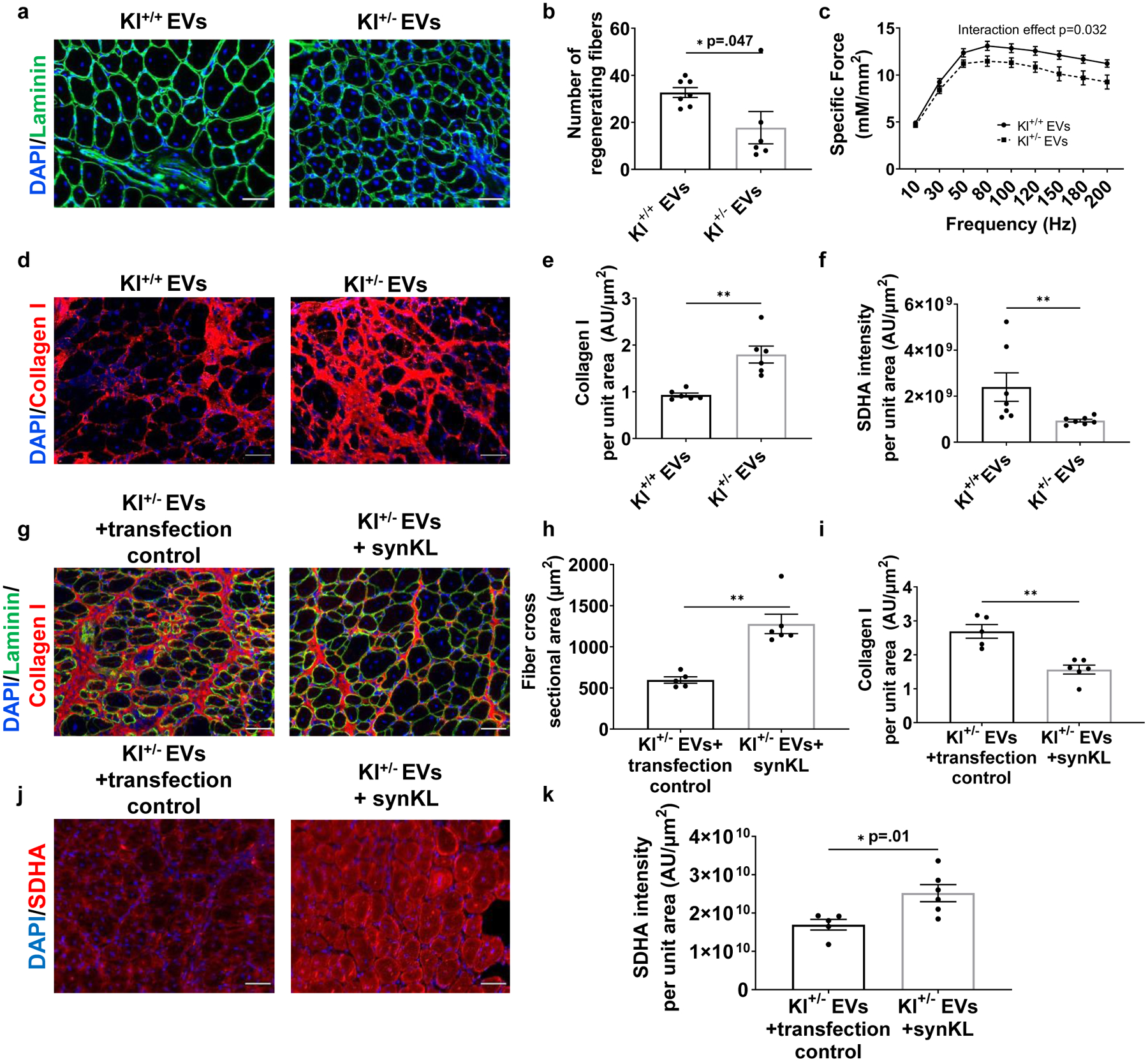
Fig. 7. Klotho mRNA within EVs contribute to functional skeletal muscle regeneration.

a, Representative images of fibers (Laminin) of injured aged TAs receiving Klotho+/+ or Klotho+/− serum EVs. Scale: 50 μm. b, Number of regenerating fibers greater than 600μm2 in injured TAs of aged mice receiving Klotho+/+ or Klotho+/− serum EVs. (*p<0.05, two-tailed Mann Whitney test, n=6 (Kl+/−), 7 (Kl+/+)). c, Specific tetanic force frequency curves of aged animals receiving EVs isolated from Klotho+/+ or Klotho+/− serum (two-way mixed ANOVA, repeated measures with frequency, interaction effect of frequency and experimental group p=0.032, n=18 (Kl+/+), 20(Kl+/−)). d, Representative images of Collagen I in injured muscles of aged mice receiving Klotho+/+ or Klotho+/− serum EVs. Scale: 50 μm. e, Quantification of Collagen I in injured muscle cross-sections of aged mice receiving Klotho+/+ or Klotho+/− serum EVs. (**p<0.01, two-tailed Welch’s t-test, n=6/group). f, Quantification of SDHA of regenerating myofibers in injured TAs of aged mice receiving Klotho+/+ or Klotho+/− serum EVs. (**p<0.01, two-tailed Mann Whitney test, n=7/group). g, Representative images and histological analysis of h, fiber cross-sectional area and i, collagen I of injured TAs of aged mice receiving Klotho+/− serum EVs loaded with transfection control or synthetic Klotho mRNA. (h: **p<0.01, two-tailed Mann Whitney test; i: **p<0.01, two-tailed Mann Whitney test; n=5 (Kl+/− EVs+transfection control), 6 (Kl+/− EVs+ synKL)). Scale: 50 μm. j, Imaging and k, quantification of SDHA in regenerating myofibers at the site of injury receiving Klotho+/− serum EVs loaded with transfection control or synthetic Klotho mRNA. (*p<0.05, two-tailed Welch’s t-test, n=5 (Kl+/− EVs+transfection control), 6 (Kl+/− EVs+ synKL)). Data presented as mean ± SEM.
