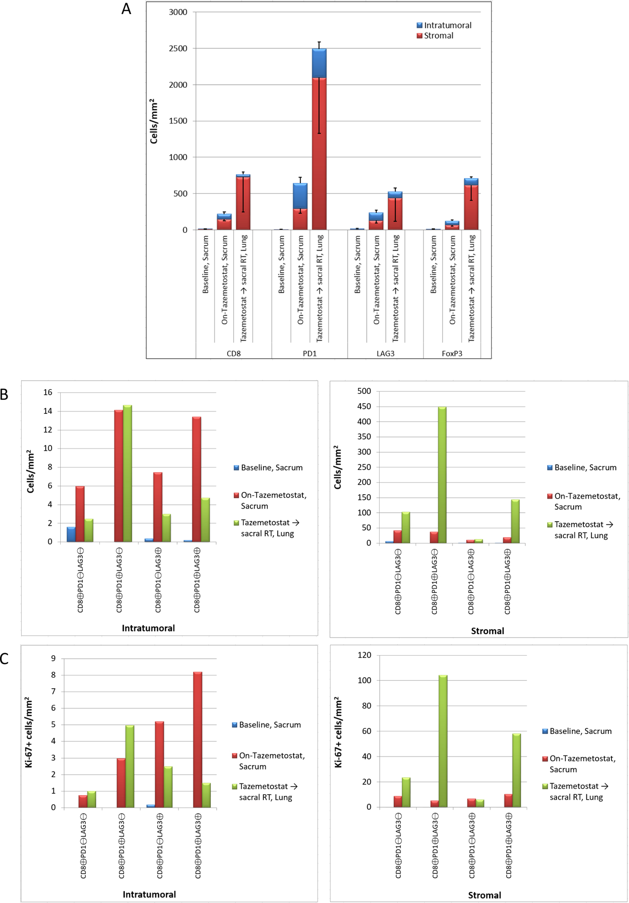
FIGURE 3.

Panel A) Comparison of intratumoral and stromal expression of immune cell markers CD8, PD-1, LAG-3, and FoxP3 in “Baseline, Sacrum”, “On-tazemetostat, sacrum”, and “Tazemetostat followed by radiation, lung” biopsy samples, quantitated as cells/mm2. Panel B) Comparison of intratumoral (left panel) and stromal (right panel) CD8+ CTL subtypes based on co-expression of PD-1 and LAG-3 in each biopsy, quantitated as cells/mm2.) Panel C) Proliferative indices of CD8+ CTL subtypes based on PD-1 and LAG-3 co-expression (B). The number of intratumoral (left panel) and stromal (right panel) Ki67-positive CD8+ CTL subtypes in each biopsy sample is plotted (cells/mm2).
