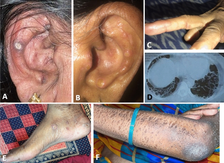
Fig. 3.
Case 2. Ulcerative lesion on right ear (A) and numerous papules seen over left antihelix (B). Distal finger with erythematous maculopapular lesion. (C). CT thorax showing ground glass opacities in bilateral lower lobes (D). Several ulcerative lesions seen on medial right foot and ankle (E). Ichthyosis over left forearm (F)

