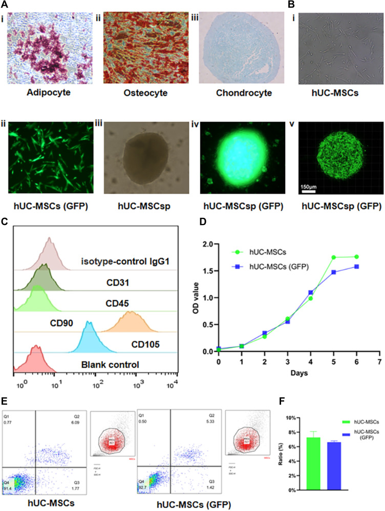
Figure 1.
Identity, transfection and spherification of hUC-MSCs. (A) Representative images (100×) of adipogenesis (i), osteogenesis (ii), and chondrogenesis (iii) of hUC-MSCs stained with oil red O, alizarin red S, and Alcian blue, respectively. (B) Transfection and spherification of hUC-MSCs were performed. hUC-MSCs (GFP) (ii) were obtained by transfecting lentiviruses with GFP into hUC-MSCs (i). hUC-MSCsp (iii), and hUC-MSCsp (GFP) (iv and v) were observed by inverted fluorescent microscopy and confocal laser scanning microscopy, respectively. (C) Surface markers of hUC-MSCs, including isotype-control IgG negative, CD31 negative, CD45 negative, CD90 positive, and CD105 positive cells, were detected by flow analysis. (D) The proliferation ability of the hUC-MSCs group and hUC-MSCs (GFP) groups was detected by CCK-8 at 1 week. (E and F) Apoptosis of hUC-MSCs, hUC-MSCs, and hUC-MSCs (GFP) groups was detected by flow analysis (annexin V-APC and 7-AAD).

