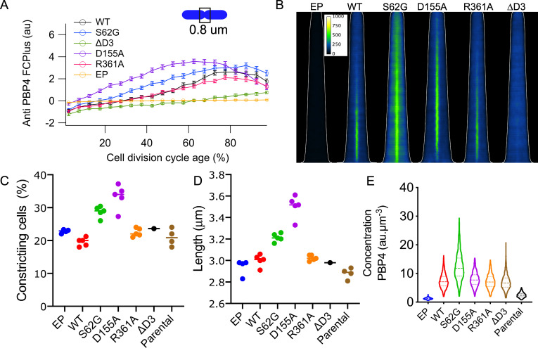
Fig 5. Localization of PBP4 depends on the presence of domain 3 but not on activity.
Cells were grown in minimal glucose medium to steady state at 28°C, fixed and immunolabeled with antibodies against PBP4. (A) The extra fluorescence at midcell (FCPlus) in the ΔdacB strain transformed with the empty plasmid (EP, yellow), or plasmids expressing wild-type PBP4 (black), PBP4S63G (blue), PBP4D155A, (purple) PBP4R361A (red), PBP4ΔD3 (green) was determined and plotted as function of the cell division cycle age as in bins of 5% age classes with the error bar indicating the 95% confidence interval. (B) Demographs of the localization fluorescence pattern of the PBP4 variants shown in (A) with the cells sorted according to their cell length. The white line indicates the length of the cells. Intensity scaling is identical for all demographs. Number of cells analyzed for each immunolabeling was at least 2000 cells. Graphs with the percentage of constricting cells (C) and the average cell length in μm (D) for the various mutants expressed from plasmid without induction in the ΔdacB strain and of the parental strain BW24113 (n = 4). (E) The concentration of PBP4 in fluorescence units per μm3 in these cells for a representative experiment (out of the four repeats).

