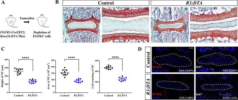
Fig. 4.
Ablation of FGFR3+ cells inhibits the postnatal development of NP tissue. (a) Schema shows the depletion of the FGFR3+ cells by Tam administration in FGFR3-CreERT2;Rosa26-DTA (R3;DTA) mice. (b) Representative Safranin O/Fast green staining images reveal pathological changes in IVD from R3;DTA mice induced with Tam at P7 and sacrificed at 2w. Scale bars, 100 µm. (c) Height, area and cell counting of NP of R3;DTA mice compared with the control FGFR3-CreERT2 after Tam administration are compared, respectively. ****P < 0.0001, Unpaired Student’s t test. (d) TUNEL assay and Ki67 immunostaining were performed to detect the changes of death and proliferation of NP (dotted circle) cells after FGFR3P·+ cell depletion. Numbers indicate the percentages of TUNEL and Ki67 positive cells over total NP cells. Scale bars, 100 µm

