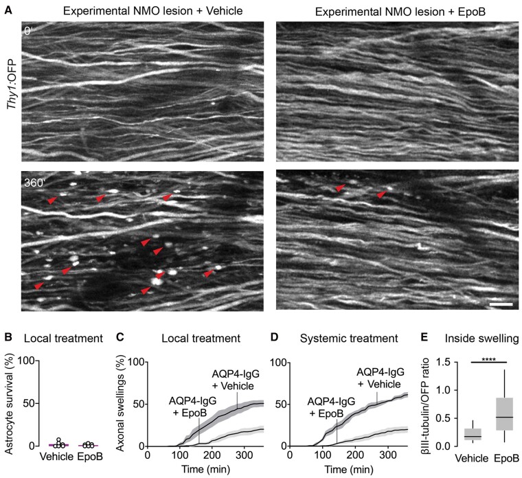
Figure 5.
Microtubule stabilization protects axons from beading. (A) In vivo two-photon imaging of Aldh1l1:GFP × Thy1:OFP mice spinal cord axons within 6 h of experimental NMO lesion induction with local epoB (5 µg/ml) or vehicle application. Red arrowheads indicate axonal beading, which is diminished with epoB treatment. Scale bar = 20 µm. (B) Unchanged depletion of spinal cord astrocytes following local epoB or vehicle treatment (survival after 3 h, epoB: 2.3 ± 1.3% versus vehicle: 1.2 ± 0.6%; n = 6 mice each). (C) Quantification of axonal beading after 6 h local epoB treatment (epoB: 19.9 ± 3.1%; vehicle: 50.9 ± 3.8%, n = 6 mice each). Mann–Whitney test, P < 0.01. (D) The fraction of beaded axons was reduced following systemic administration of epoB 24 h before lesion induction (after 6 h: 19.2 ± 3.4% versus vehicle: 61.6 ± 2.5%, n = 5, 4 mice, respectively) Mann–Whitney test; P < 0.05. (E) Quantification of βIII-tubulin staining mean fluorescent intensity (MFI) normalized to OFP signal in Thy1:OFP mice. Local epoB treatment preserved tubulin staining compared to vehicle (median, vehicle: 0.17 versus epoB: 0.52, n = 188, 65 axons respectively in five mice each). Mann–Whitney test; ****P < 0.0001. Box-and-whisker plot: 10–90 percentile. Data represent mean ± SEM in B–D.

