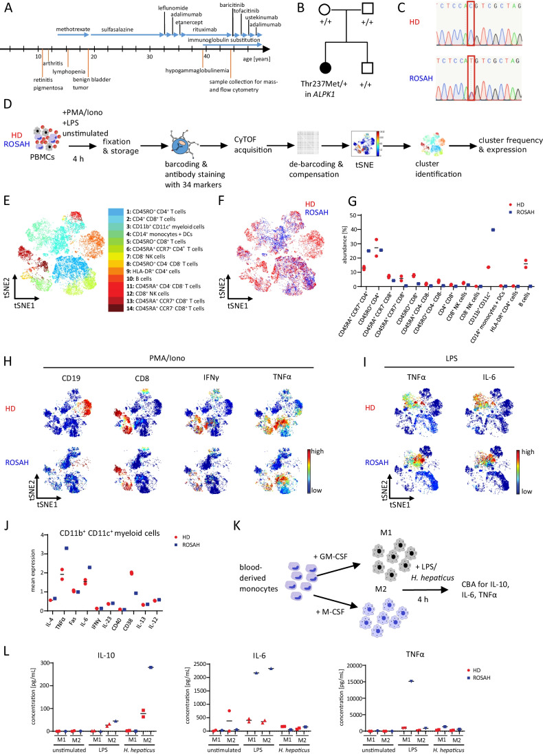
Fig. 1.
Deep immune phenotyping of a patient with chronic auto-inflammation and progressive vision loss reveals an increased expression of TNFα and IL-6 in myeloid cells. A Summary of the clinical history of the ROSAH patient. B Variant status in the ALKP1 gene in the ROSAH patient, her parents, and her brother as assessed by whole-exome sequencing, “ + ” indicating the wild type allele, Thr237Met indicating the mutated allele with missense mutation (p.Thr237Met). C Sanger sequencing of EDTA blood of the ROSAH patient and an unrelated healthy donor (HD). The red square indicates the position of the mutation (c.710C) in the ALPK1 gene. D Schematic summary of immune cell characterization of the ROSAH patient and two unrelated HDs by mass cytometry. Peripheral blood mononuclear cells (PBMCs) of the ROSAH patient and HDs were ex vivo stimulated with phorbol 12-myristate 13-acetate (PMA)/ionomycin (Iono) or lipopolysaccharide (LPS) for 4 h followed by fixation and mass cytometry staining and acquisition. E t-SNE plot of concatenated FCS files from all samples, colored by the 14 clusters identified in CD45+ cells. F t-SNE plot displaying the cellular distribution of CD45+ cells of the ROSAH sample (blue) and two HD samples (red). G Abundance of the 14 different cell clusters in PMA/Iono-stimulated PBMCs. H t-SNE plots of PMA/Iono-stimulated PBMCs colored by the expression of selected markers. Red represents high expression; blue represents low expression. I t-SNE plots of LPS-stimulated PBMCs colored by the expression of selected markers. Red represents high expression; blue represents low expression. J Mean expression of selected markers in CD11b+CD11c+ myeloid cells of LPS-stimulated PBMCs. K Blood-derived monocytes of the ROSAH patient and one HD were differentiated into M1 or M2 macrophages by supplementation of GM-CSF or M-CSF for 7 days and subsequently stimulated with LPS or Helicobacter hepaticus (H. hepaticus). L Concentrations of IL-10, TNFα, and IL-6 in the supernatant of stimulated macrophages. Duplicates represent two wells of separately differentiated macrophages from the same HD

