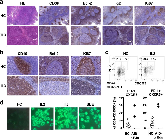
Fig. 2.
Exaggerated germinal center activity in AID-ΔE4a patients. a Representative histological (HE staining) and immunohistological (CD38, Bcl-2, IgD, Ki-67) analysis of tonsil tissue derived from patient II.3 and a control individual (magnification × 100). b Immunohistological analysis (CD10, Bcl-2 and Ki67) of germinal centers (magnification × 400). c Representative dot plot of PD-1 and CXCR5 surface expression on peripheral blood CD4+ CD45RO+ T cells (upper part) and frequencies of TFH (PD-1+CXCR5+CD45RO+CD4+) and TPH (PD-1+ CXCR5−CD45RO+CD4+) cells in AID-ΔE4a patients and age-matched healthy controls as assessed by flow cytometry (lower part). d Indirect immunofluorescence staining of HEp-2 cells using FITC-labeled secondary anti-IgM-antibodies with indicated sera derived from both AID-ΔE4a patients, a SLE patient and a healthy control (dilution 1:160)

