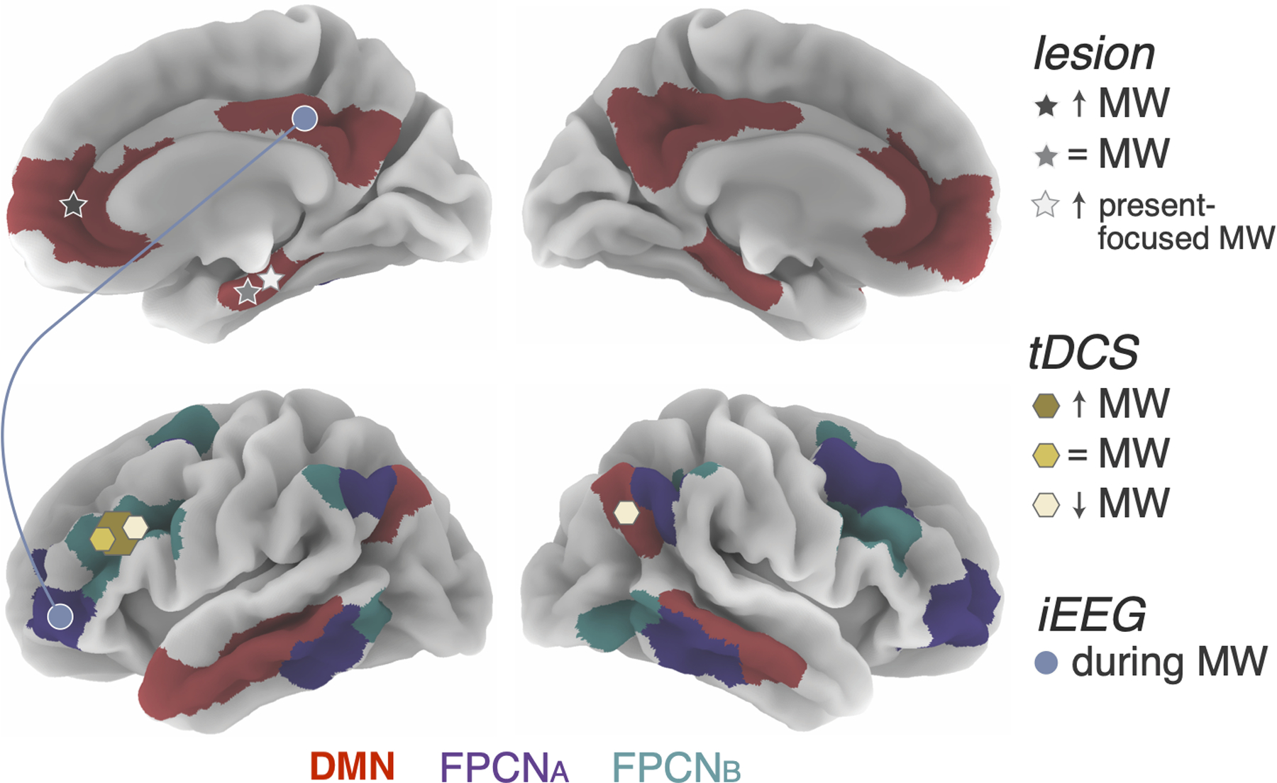
Figure 1. Graphical summary of the role of DMN and FPCN subregions in mind wandering.

The effects of lesion on mind-wandering frequency and temporal focus of mind-wanderirng are indexed by star shapes. Darker grey indicates a lesion in that location was shown to increase mind-wandering frequency (primarily in the ventromedial PFC); the medium shade of grey indicates an absence of significant effect on mind-wandering frequency (in the hippocampus); the lightest grey indicates an increase in present-focused thoughts during MW (in the hippocampus). tDCS effects primarily focusing on mind-wandering frequency are indexed by hexagonal shapes: darker yellow indicates stimulation of that region was shown to increase mind-wandering (in the DLPFC), lighter yellow indicates a decrease in mind-wandering (in the DLPFC and right IPL), and the medium shade indicates an absence of significant effect (in the DLPFC). The size of the hexagon corresponds to the proportion of papers reporting that effect; however, note the sample size across studies is not reflected in this figure. Connectivity between DMN and FPCN subsystem A during mind-wandering as revealed by iEEG studies are shown in blue circles, which reflects involvement of the entire network in which the circle is located.
