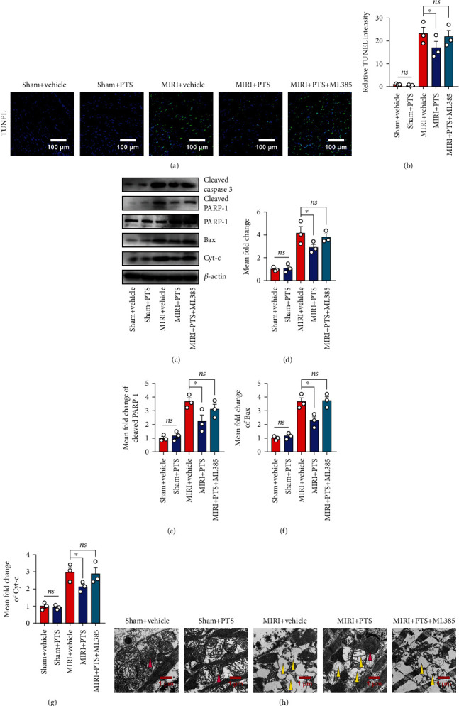
Figure 7.

PTS ameliorated myocardial mitochondrial apoptosis in MIRI rats by regulating Nrf-2. A total of 50 rats were divided into five groups to explore the underlying mechanisms including the I/R group, sham group, sham+PTS group, I/R+PTS group, and PTS+ML385 group. (a) Representative TUNEL results of myocardial tissue sections. (b) The relative TUNEL staining intensities of each group were quantified using ImageJ software. (c) Western blotting analysis showed the changes in apoptosis-related proteins in myocardial tissue after Nrf-2 blockade. (d–g) The levels of cleaved caspase-3, cleaved PRAR-1, Bax, and Cyt-c were quantified using ImageJ. (h) The ultrastructure of the rat myocardium was examined by transmission electron microscopy. The red arrow indicates normal myocardial mitochondria, and the yellow arrow indicates damaged mitochondria. Data are expressed as the mean ± SEM (n = 3). ∗P < 0.05, ∗∗P < 0.01; ns: not significant.
