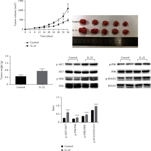
Figure 7.

IL-22 accelerated the tumor growth in the xenograft models. A549 cells were subcutaneously injected into the right armpit of BALB/c nude mice. When the tumor volumes almost reached 50-100 mm3, these mice were randomly divided into two groups. Each group of mice was injected with IL-22 (4 μg, daily; five days per week) or vehicle alone (n = 5). (a) The tumor volumes of mice were determined every three days after the onset of treatment. (b, c) Finally, these mice were sacrificed under anesthesia. Then, the entire tumor was resected, and the weight of the tumor was measured. (d, e) Subsequently, the harvested tumors were lysed, and western blot analysis was performed for p-AKT, AKT, p-p38, p38, p-ERK, ERK, p-STAT3, and STAT3. The data were presented as mean ± standard deviation (SD). The significant differences were compared with the controls and were indicated as ∗P < 0.05, ∗∗P < 0.01, and ∗∗∗P < 0.001.
