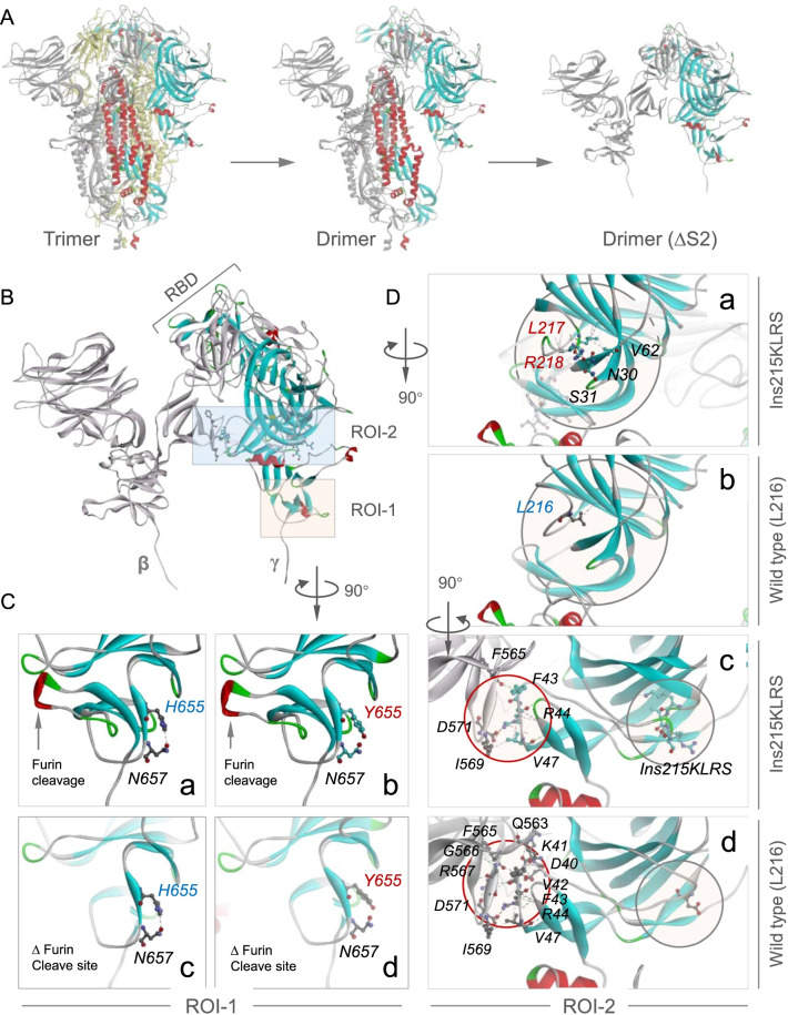
Fig. 3.
Three-dimensional structure modeling of the MA-SARS2 spike. A Side view of S glycoprotein whose chain colored in Yellow/ Gray/ Rainbow, Drimer (ΔS2): two protomers lacking S2 subunit. B Zoomed-in of Drimer (ΔS2) as indicated. ROI-1: region 1 of interest, ROI-2: region 2 of interest; β, γ: S1 subunit of S glycoprotein. C Zoomed-in of ROI-1 after clockwise rotation by 90 degrees. Images show the interactions between H655 (a, c) or Y655 (b, d) with N657 in the presence or absence of furin cleavage finger. D Zoomed images of ROI-2 after clockwise flipping by 90 degrees (a, b) and subsequent clockwise rotation by 90 degrees (c, d). Images a, b show the interactions between two chains, c, d show the interactions between wild type (L216) or Ins215KLRS and other amino acids. Gray circle: Ins215KLRS mutation

