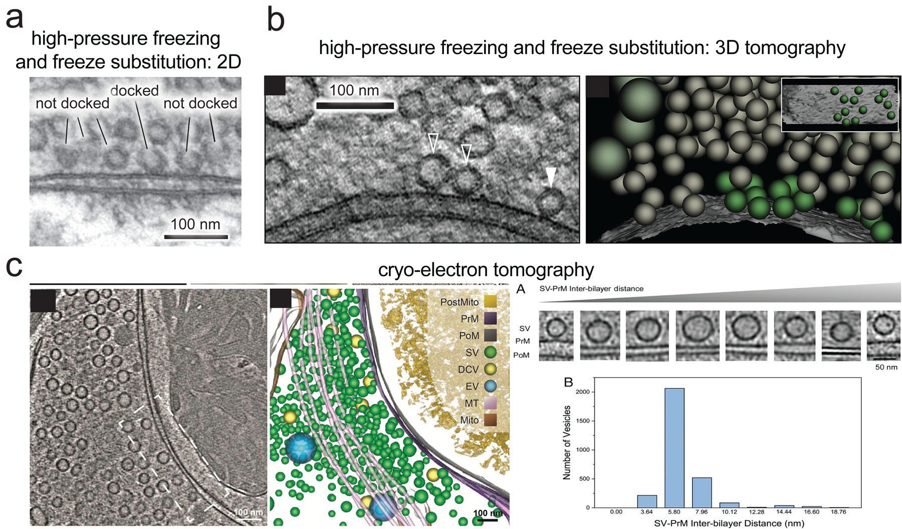
Figure 1.

The strict definition of synaptic vesicle docking.
What is meant by ‘docking’ in synaptic ultrastructure varies from study to study. This term is often used for all vesicles within 30–40 nm of the plasma membrane at the active zone (measuring the nearest distance between the edge of the vesicle membrane and plasma membrane). Here, we refer to docking by a strict definition: structurally, docking is the closest synaptic vesicles can get to the plasma membrane at the active zone before fusion as observed by electron microscopy. (a) In high-pressure frozen and freeze-substituted samples, docked vesicles make a ‘point contact’ with the plasma membrane, visible in both (a) 2D thin sectioning EM and (b) 3D electron tomography (solid arrowheads indicate vesicles with visible plasma membrane contact in the tomograph slice shown, hollow arrowheads indicate vesicles that are docked and make contact with the plasma membrane, but the contact is not visible in this slice; green vesicles in the 3D rendering are docked), with no apparent space between vesicle membrane and plasma membrane down to the effective resolution of this technique (0–2 nm) [28,43]. (c) In cryo-electron tomography, which visualizes the native state of tissue under vitreous ice without any staining, dehydration, or fixation, the closest vesicles get to the plasma membrane in synapses at rest is ~5 nm [84–86], and by our definition these constitute docked vesicles. This means the apparent 0–2 nm distance in freeze substituted samples is likely an artifact. However, the two characteristic distances in these techniques are likely both meaningful and correspond to the same vesicles. ~75% of all vesicles within 20 nm of the plasma membrane accumulate at this closest distance in cultured hippocampal synapses, regardless of which technique is used [28,84]. [27,82]. Accumulation at this specific distance is unique to docking, as undocked vesicles within 100 nm are roughly evenly distributed in distance from the active zone. Only this closest stage of approach requires SNARE complex assembly [27,43], only docked vesicles are depleted by stimulation [26,28], and in cryo-electron tomography only these vesicles are connected to the membrane by a stereotyped protein density that may correspond to the docking/fusion machinery [84]. All these lines of evidence together strongly argue that docked vesicles, and only docked vesicles, are at the final stage of priming and readiness for fusion. Vesicles that are close to the plasma membrane, but not docked, we refer to simply as undocked or as ‘replacement vesicles’ (these vesicles are sometimes referred to as ‘tethered’). In terms of distance from the plasma membrane by EM, our definition of docking corresponds to the term ‘tightly docked’ often used in the field [7]. Note that any studies using traditional chemical fixation for electron microscopy, rather than fast freezing, cannot resolve the distinctions discussed here. Aldehyde fixation of living tissue causes severe deformations in cellular structures [87] and directly triggers synaptic vesicle exocytosis [88], making evaluation of fine structure near the active zone inaccurate. For example, under chemical fixation, preventing SNARE complex assembly has no apparent effect on docking [89].
(a) and (c) are reproduced, with permission, from [43] and [84], respectively. (b) is reproduced from [26].
