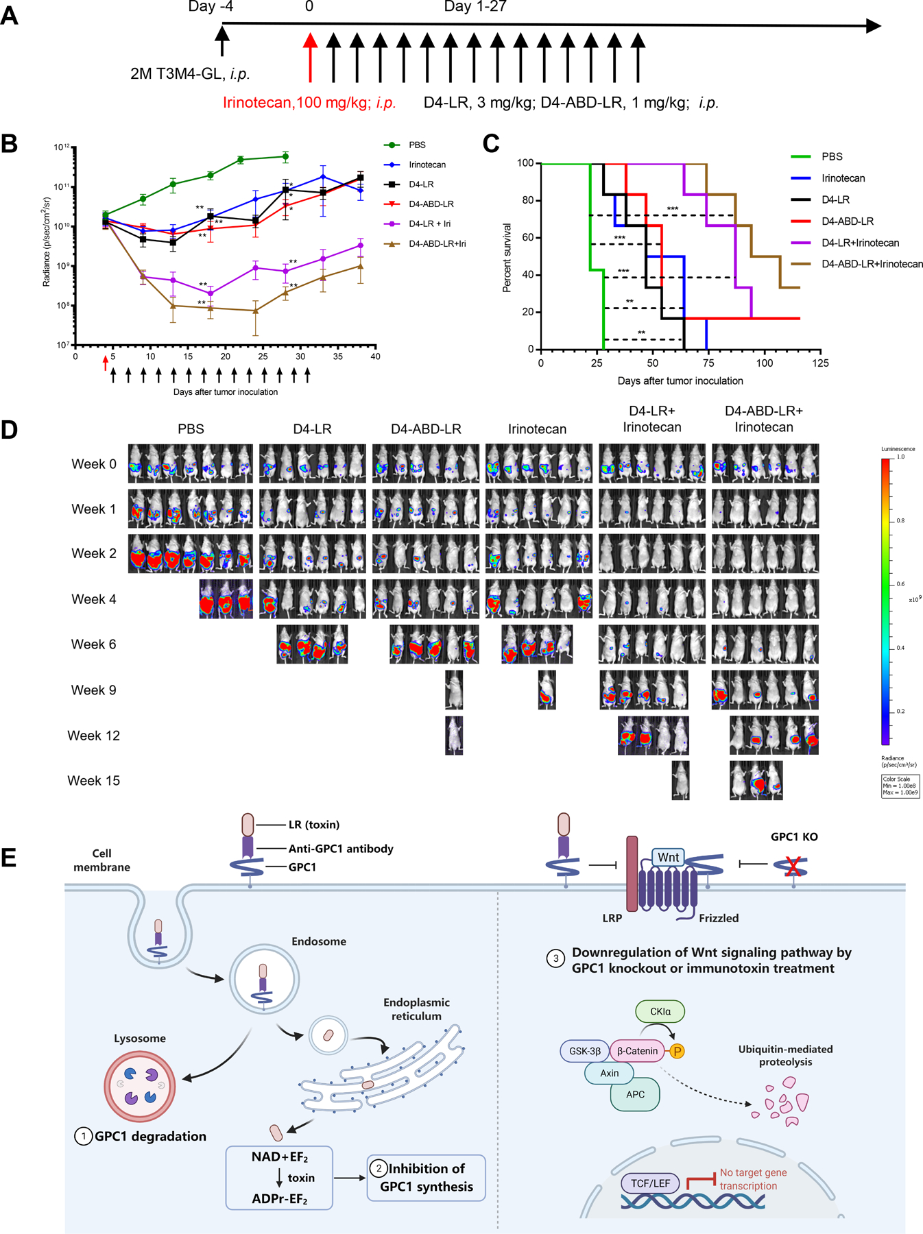
Figure 6. D4-ABD-LR in combination with irinotecan showed enhanced anti-tumor efficacy.

A Timeline of the T3M4 mouse model treated with D4-LR or D4-ABD-LR as a single agent or combined with irinotecan. Six-week-old female athymic nude mice were intraperitoneally injected with 2 × 10⁶ T3M4 cells. The red arrow represents the administration of irinotecan at 100 mg/kg. D4-LR at 3 mg/kg or D4-ABD-LR at 1 mg/kg was intraperitoneally injected into mice on the days indicated by black arrows. N=7 for PBS control group, n=6 for other treating groups. B, Average tumor volume ± SEM for each group. C, Kaplan-Meier survival curve. D, Bioluminescent imaging showing tumor burden of an individual mouse. E, A schematic describing three potential mechanisms employed by anti-GPC1 immunotoxins to inhibit pancreatic tumor cells growth. Upon binding to GPC1 on the tumor cell surface, the GPC1/immunotoxin complex is internalized by endocytosis. In the endosome, the immunotoxin is processed by the protease furin to separate the antibody fragment from the toxin. The antibody fragment together with GPC1 goes to the lysosome where it is degraded. In contrast, the toxin is transferred to the endoplasmic reticulum and then enters the cytosol. After reaching the cytosol, the toxin resulted in the inhibition of GPC1 synthesis via mediating ADP-ribosylation of EF2. In addition, GPC1 knockout or immunotoxin treatment reduces Wnt binding to Frizzed/LRP, leading to the downregulation of Wnt/β-catenin signaling. Abbreviations: ADPr, ADP-ribose; APC, adenomatous polyposis coli; CKIα, casein kinase Iα; EF2, elongation factor 2; GSK-3β, glycogen synthase kinase-3β; KO, knockout; LRP, low-density lipoprotein receptor-related protein; NAD, nicotinamide adenine dinucleotide; TCF/LEF, T cell factor/lymphoid enhancer factor. Figure 6E was created with BioRender (https://app.biorender.com).
