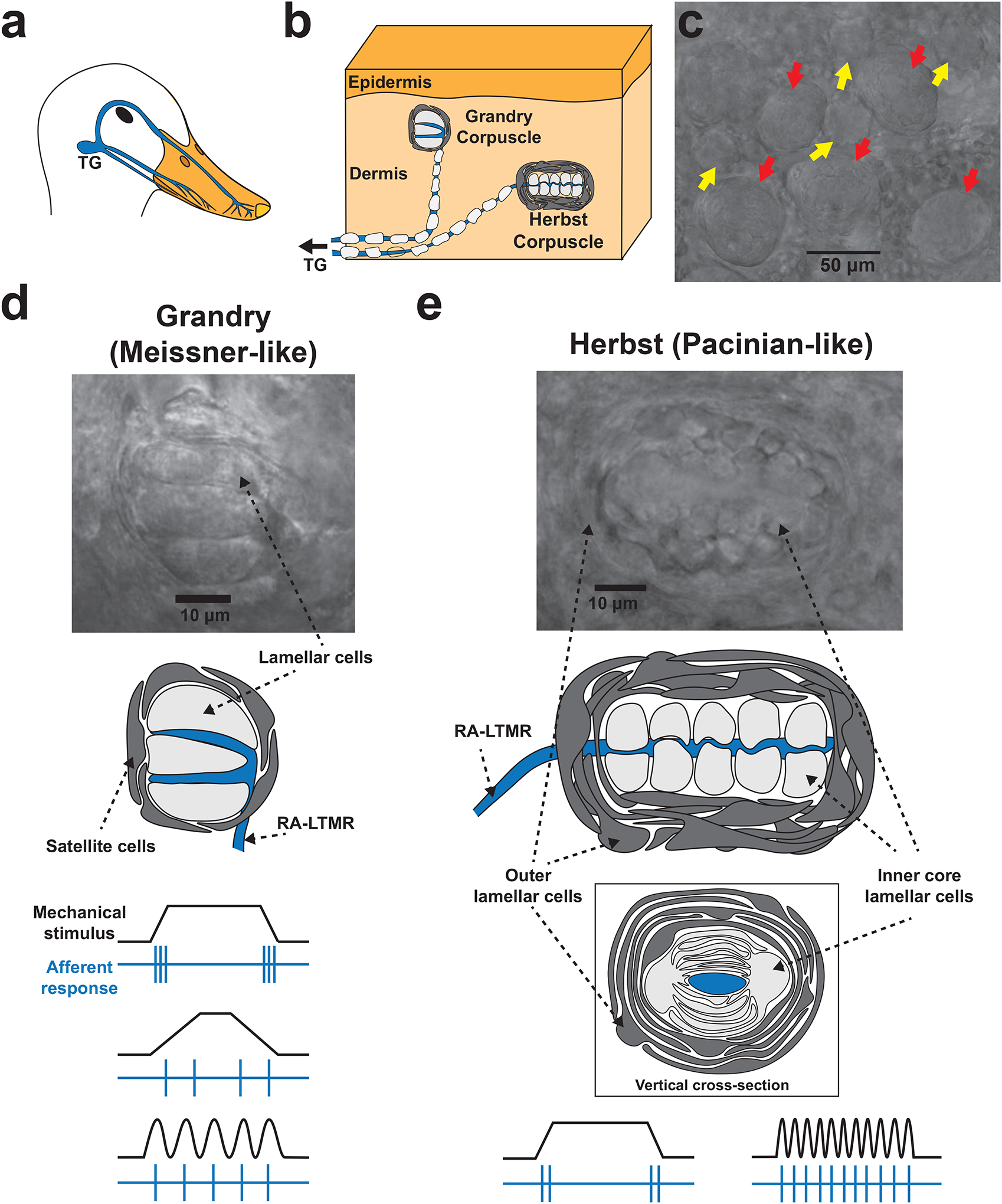
Figure 1. The structure and function of Grandry and Herbst corpuscles within the mechanosensory bill of waterfowl.

(a) The neuroanatomy underlying tactile sensation in the bill. LTMRs from the trigeminal ganglia (TG) project to the skin of the bill. (b) Trigeminal LTMRs form terminal end-organs in the bill dermis, most of which are Grandry and Herbst corpuscles. (c) Image of the bill dermis under a brightfield microscope. Grandry (yellow arrows) and Herbst (red arrows) corpuscles are present at a cumulative density of up to 200 corpuscles per square millimeter of skin, and can be easily distinguished by size and morphology. (d) Higher magnification image and diagram of a Grandry corpuscle. The Grandry corpuscle is composed of 2–12 lamellar cells which are layered above and below the terminals of the RA-LTMR. The structure is encapsulated by satellite cells. Below the diagram, example stimuli (black) and LTMR afferent responses (blue) are shown. The LTMR of the Grandry corpuscle detects changes in transient force, low frequency vibration, and velocity; the impulses/second of the LTMR response increase with increasing velocity of the mechanical stimulus. (e) Higher magnification image and diagram of a Herbst corpuscle. The Herbst corpuscle is composed of an outer capsule formed by outer core lamellar cells, which encloses an inner core comprised of inner core lamellar cells. The outer core and inner core lamellar cells form concentric lamellae surrounding the mechanoreceptor afferent. Below the diagram, example stimuli (black) and LTMR afferent responses (blue) are shown. The LTMR of Herbst corpuscles detects transient force and high frequency vibration.
