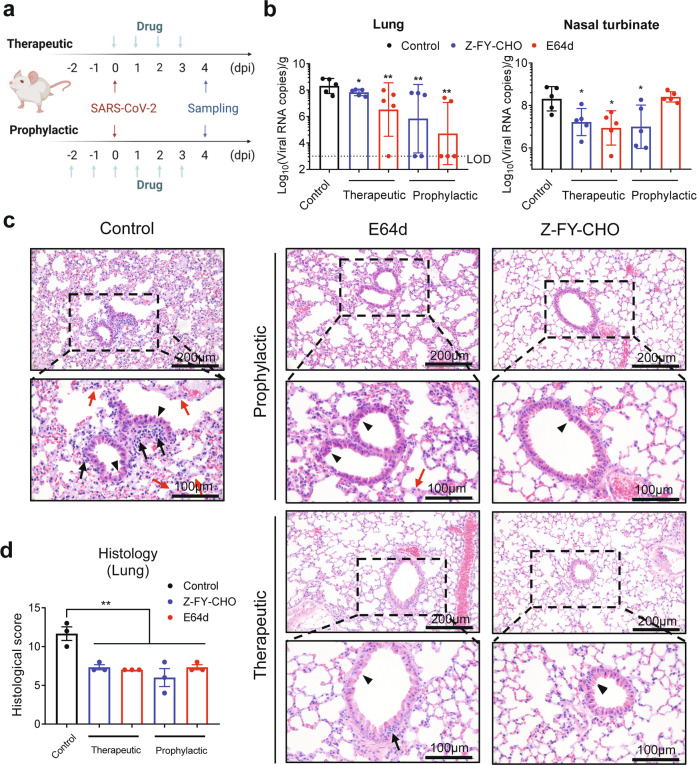
Fig. 5. CTSL inhibitors prevent SARS-CoV-2 infection in vivo.
a E64d and Z-FY-CHO were administered intraperitoneally at 2 days before infection to 3 dpi as prophylactic treatment, and mice were challenged with 106 PFU at 0 dpi; the two drugs were administered therapeutically at 0–3 dpi. Tissue samples were collected at 4 dpi. b Viral RNA copies in mouse lung and nasal turbinate tissues (n = 5 mice/group). The dotted line indicates the lower limit of detection (LOD). Statistical significance was assessed between the indicated group and control group by one-way ANOVA with Tukey’s post-hoc test. c Representative images from histological analysis of lungs from SARS-CoV-2-infected hACE2 mice at 4 dpi. Magnified views of the boxed regions in each image are shown below the corresponding image. The black arrows indicate inflammatory cell infiltration, the black arrowheads indicate bronchiolar epithelial cell degeneration, and the red arrows indicate alveolar septal thickening. The scale bars are indicated in the figures. d Semiquantitative histological scoring of each lung tissue was performed by grading the severity of bronchiolar epithelial cell damage (0–10), alveolar damage (0–10) and inflammatory cell infiltration in blood vessels and bronchioles (0–10) and summing these scores to calculate the total score. Normal = 0, indeterminate = 1–2, mild = 3–4, moderate = 5–7, severe = 8–10. (n = 3) Statistical significance was assessed between the indicated group and control group by one-way ANOVA with Tukey’s post-hoc test.

