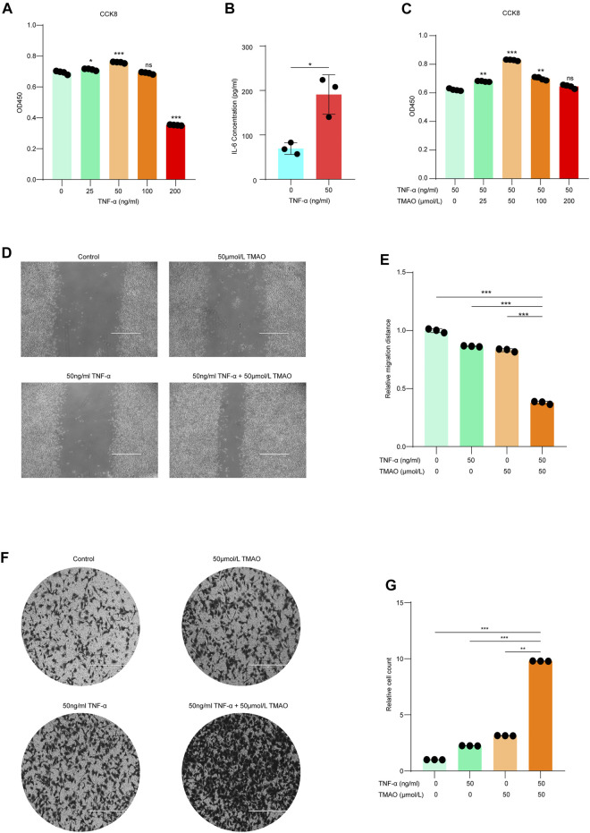
FIGURE 1.
TMAO synergistically enhances the tumorigenicity of Hepa1-6 cells in the presence of TNF-α. (A) TNF-α dose-dependently promoted the proliferation of Hepa1-6 cells; maximum proliferation was observed at 50 ng/ml TNF-α; results are mean ± SD (n = 3 per group); one-way ANOVA with Bonferroni’s test; ***, p < 0.001. (B) Hepa1-6 cells treated with 50 ng/ml TNF-α expressed higher levels of IL-6 compared to untreated cells; results are mean ± SD (n = 3 per group); Student’s t-test; ***, p < 0.001. (C) The combination of 50 ng/ml TNF-α and 50 μM TMAO led to the maximal synergistic increase in cell proliferation; results are mean ± SD (n = 3 per group); one-way ANOVA with Bonferroni’s test; ***, p < 0.001. (D,E) Wound healing assay confirmed that 50 ng/ml TNF-α alone and 50 μM TMAO alone slightly promoted the migration of Hepa1-6 cells compared to untreated control cells, while the combination of 50 ng/ml TNF-α and 50 μM TMAO synergistically and significantly promoted the migration of Hepa1-6 cells. Scale bars = 1,000 μm; data were analyzed by ImageJ; data are mean ± SD (n = 3 per group); one-way ANOVA with Bonferroni’s test. (F, G) Migration assay confirmed that 50 ng/ml TNF-α alone and 50 μM TMAO alone slightly promoted the migration of Hepa1-6 cells compared to untreated control cells, while the combination of 50 ng/ml TNF-α and 50 μM TMAO synergistically and significantly promoted the migration of Hepa1-6 cells. Scale bars = 400 μm; data were analyzed by ImageJ; data are mean ± SD (n = 3 per group); one-way ANOVA with Bonferroni’s test.

