Fig. 4.
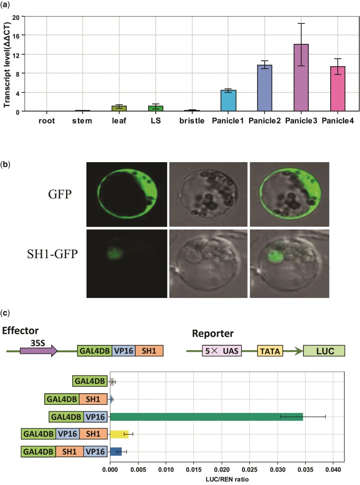
Gene function analysis of sh1 in foxtail millet. (a) Expression levels of sh1 in multiple organs including the root, stem, leaf, leaf sheath (LS), bristle, and panicles at different stages (Panicle1, before heading; Panicle2, 3 d after heading; Panicle3, 8 d after heading; Panicle4, grain-filling stage). (b) Subcellular localization of the SH1–GFP fusion protein in foxtail millet leaf cells. (c) Dual-luciferase transient activity assays determined that SH1 functioned as a transcriptional repressor. The GAL4DB–VP16–SH1 fusion protein dramatically (P = 6.9 × 10⁻⁵) repressed luciferase activity in comparison to the control protein GAL4DB–VP16. **, strongly significant; error bars, SD (n = 3).

