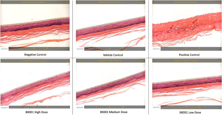
FIGURE 4.

Histopathological evaluation of reconstituted human epithelial tissues exposed to BX001 phages at different doses. Tissues were sectioned at approximately 5 μm thickness and stained with haematoxylin and eosin. No treatment‐related changes were seen in the negative control, vehicle control or any sample exposed to the BX001 phage preparations. Diffuse epidermal necrosis was observed in the positive control (red arrows)
