
Figure 1:
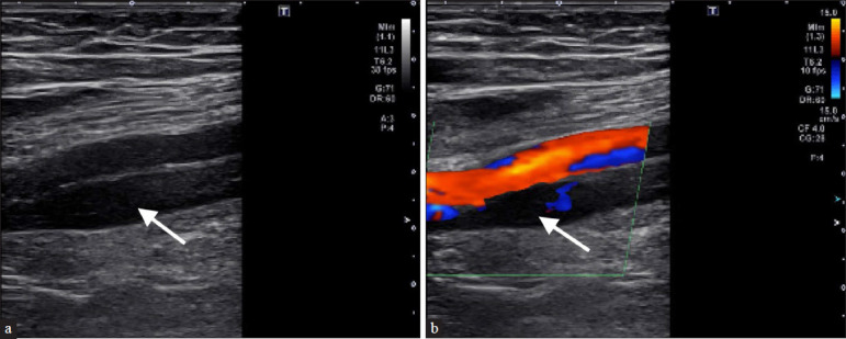
A 75-year-old male with a history of pulmonary embolism (PE) presents with lower GI bleed. Anticoagulation was subsequently discontinued, and the patient began experiencing right lower extremity pain. (a) Ultrasound (US) venous duplex demonstrates non-echogenic thrombus within the superficial femoral vein (arrow). (b) Demonstrates partial compressibility, and lack of flow on doppler imaging (arrow) consistent with acute DVT. The patient underwent temporary IVC filter placement.