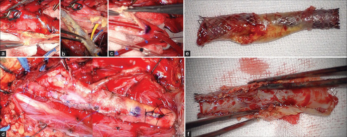
Figure 4:
(a and b) The clear boundary between the plaque, including the stent, and the vessel wall is shown. Normal carotid endarterectomy was performed. (c) Insertion of an internal shunt after removed plaque is shown. (d) The vessel wall could be sutured as normal. (e and f) The stent and plaque are shown. Neointima formed inside the lumen of the stent, causing in-stent restenosis.

