Fig. 1.
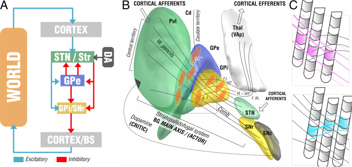
Functional-anatomical model leading to the core hypothesis for the present study. (A) Basal-ganglia model in context of a reinforcement-learning context. The left side shows the main axis of the basal ganglia (actor) with a three-layer model in which both striatum and subthalamic nucleus form entry nodes and GPi and substantia nigra pars reticularis (SNr) serve as output ganglia, feeding information back (via the thalamus) to the cortex and passing it on to brainstem centers (BS). Dopaminergic input serves as one of multiple critics to reinforce successful motor behavior. Adapted from ref. 16. (B) Translation of the model to the anatomical domain based on information shown in SI Appendix, Fig. S1. The striatopallidofugal system and pallidothalamic fibers serve as the main axis (actor) and receive feedback from dopaminergic centers, especially the substantia nigra pars compacta (SNc). Pallidal receptive fields reside in a 90° angle to the striatopallidofugal fiber system, pallidothalamic output tracts traverse the main axis in equally orthogonal fashion. (C) Hypothesis generation for the present study based on anatomical considerations. Two scenarios are possible (shown as cut-out box from B). (Upper) Active contacts (pink) of top-responding patients are located along the direction of striatopallidonigral fibers. In this case, our results would reveal activation of these fibers to best account for clinical outcome. (Lower) Instead, active contacts (cyan) could also be located along pallidothalamic tracts (ansa lenticularis; a.l. and fasciculus lenticularis; f.l.). In this case, our results would reveal activation of these fibers to best account for clinical outcomes.

