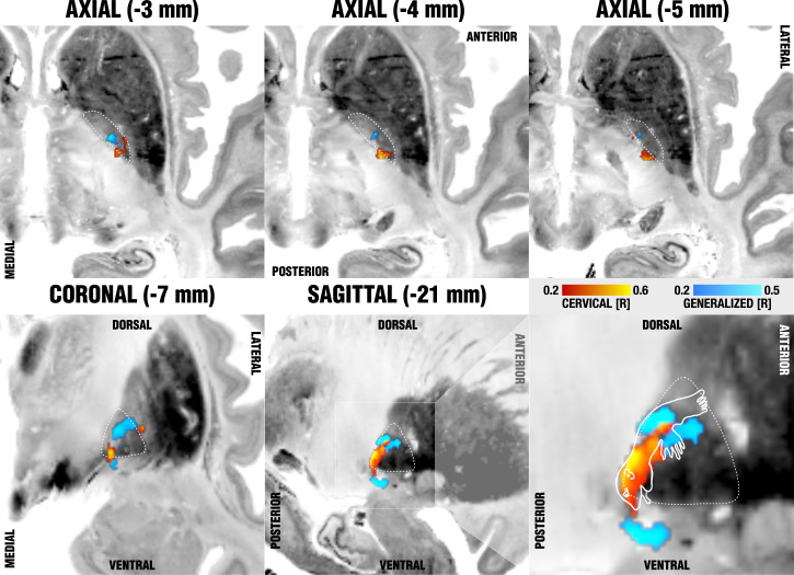
Fig. 4.
Sweetspot mapping of cervical (red) vs. generalized (blue) subcohorts matches somatotopic organization of the GPi as defined by Nambu (12). Voxels are color-coded by the degree of correlation between percent improvements of either TWSTRS (cervical, hot colors) or BFMDRS (generalized, cool colors) and shown on multiple axial (Upper) and coronal/sagittal (Lower) slides on top of the BigBrain template (50). The last panel (Lower Right) shows the homuncular representation of the pallidum following reports by Nambu (12), which stated that neurons responding to the orofacial, forelimb, and hindlimb regions of motor cortex are located along the ventral-to-dorsal axis in the GPi.

