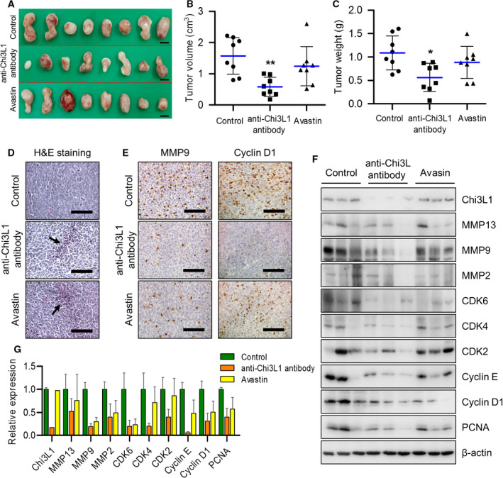
Fig. 2.

Anti‐Chi3L1 antibody blocks tumor growth in vivo. (A–C) Lewis lung cancer (LLC) cells (3 × 10⁵ cells) were injected subcutaneously to induce lung cancer tumors and 0.5 mg·kg−1 of vehicle, anti‐Chi3L1 antibody, or Avastin were intravenously administrered twice a week for 4 weeks. n = 8 per group. (A) Representative image of tumors obtained from mice in each group. Scale bar, 1.5 cm. (B, C) Tumor size and weight were measured, and tumor volume was calculated with the formula; V = L × W2 × 0.5 (n = 8). *P < 0.05; **P < 0.01; (one‐way ANOVA). (D) H&E staining images of tumor tissues were excised from each group. Arrows indicate apoptotic cells. H&E staining were repeated from three independent experiments. Scale bar, 400 μm. (E) Representative immunohistochemical images of tumor tissues using anti‐MMP9 and anticyclin D1 antibodies in each group. Scale bar, 100 μm. (F) The tumor tissue extracts were subjected to immunoblot analysis with indicated antibodies. (G) The intensity of each band in (F) was measured and the ratio of the amount of each protein to β‐actin was calculated. Data are presented as mean ± standard deviation (SD) from two independent experiments.
