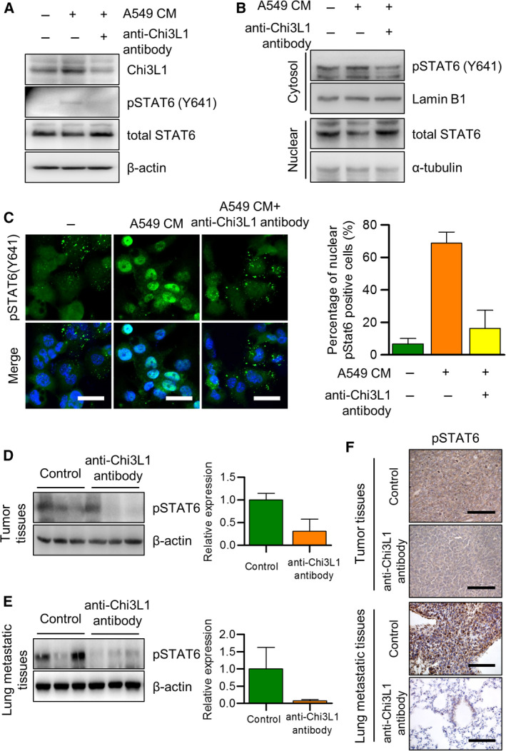
Fig. 6.

STAT6 is involved in the anti‐Chi3L1 antibody‐induced inhibition of M2‐like macrophage polarization. (A–C) THP‐1 was treated with phorbol 12‐myristate 13‐acetate (PMA) (100 ng·mL−1) and stimulated with A549 conditioned medium (CM) with or without anti‐Chi3L1 antibody (1 μg·mL−1). (A) Western blot was performed to measure the p‐STAT6. (B) Immunoblot of protein expression in cytoplasmic and nuclear fractions of THP‐1. Lamin B1 and α‐tubulin are the loading controls for nuclear and cytoplasmic fractions, respectively. (C) The fixed cells were immunofluorescence stained with p‐STAT6. The percentage of nuclear localized p‐STAT6 cells was calculated. Data are presented as mean ± standard deviation (SD) from three independent experiments. Scale bar, 20 μm. (D, E) The tumor and lung metastatic tissue samples were analyzed with immuno‐blotting with the p‐STAT6 antibody. Data are presented as mean ± standard deviation (SD) from three independent experiments. (F) The tumor and lung tissue samples were analyzed with immunohistochemistry with the p‐STAT6 antibody. Immunohistochemical staining were repeated from three independent experiments. Scale bar, 100 μm.
