Fig. 5.

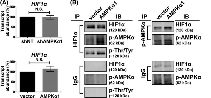
Transcriptional and post‐translational analysis of HIF1α. (A) PLC5 cells were transfected with corresponding vectors, AMPKα1, or shAMPKα1 for 72 h, and the HIF1A expression was analyzed by qPCR. Columns, mean; error bars, SD (n = 3), N.S., not significant (Welch’s t‐test). (B) PLC5 cells were transfected with the corresponding vector and AMPKα1 for 72 h. Cell lysates were collected for the immunoprecipitation assay. IgG antibody was used as a negative control of immunoprecipitation assay. The experiments were performed in three independent replicates.
