
Fig. 2.
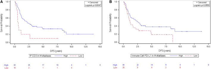
Kaplan–Meier curves showing the association between CD3high T cell and PD‐L1 markers and DFS in patients with mCRC. (A) High and low CD3 in the invasive front at the metastatic site, (B) high and low PD‐L1 expression on immune cells at the metastatic site.