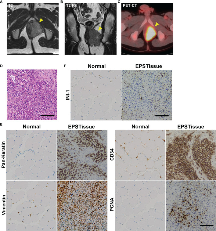
Figure 1.
Clinical images of the EPS tumor in the left perineum of a 22-year-old man. (A) T2-weighted axial image of MRI. (B) T2-weighted fat-suppressed sequence (T2 FS) coronal image of MRI. (C) The axial image of positron emission tomography-computed tomography (PET-CT). (D) Histological appearance of the original tumor with H&E staining. (E) Immunohistochemical reactivity in the original tumor. Tumor cells were diffusely positive for AE1/AE3, vimentin and CD34, but negative for INI-1 (F). MIB-1 labeling index was approximately 20%. Scale bars: 100μm.

