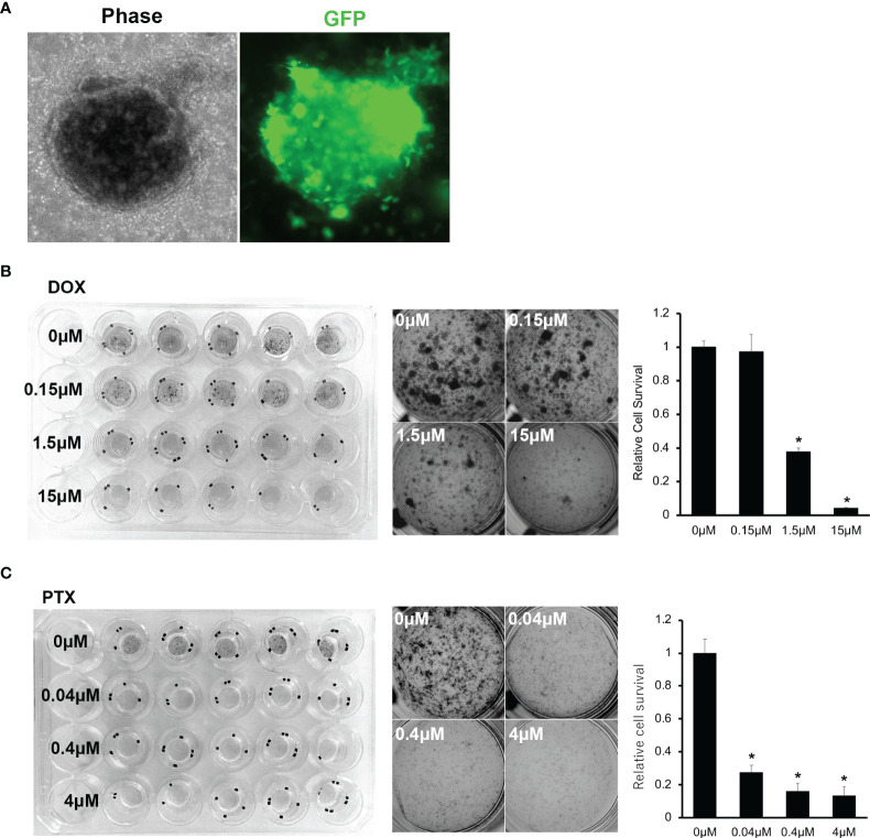
Figure 6.
(A) Microscopic images of PDO of OICI-EPS-0530 expressing green fluorescent protein (GFP). Left; OICI-EPS-0530 organoids under ALI organoid culture by phase-contrast microscopy. Right; an image of the same organoid with GFP. (B) Organoid proliferation assay with MTT assay against Doxorubicin (DOX). Left; Image of whole culture plate. Middle; Each well by concentration of DOX. Right; Relative organoids viability by concentration of DOX. (n = 5; *P < 0.005). (C) Organoid proliferation assay with MTT assay against Paclitaxel (PTX). Left; Image of whole culture plate. Middle; Each well by concentration of PTX. Right; Relative organoids viability by concentration of PTX. (n = 5; *P < 0.005).

